FotoFinder aesthetics
Systems for diagnostics, photo documentation, and consultations in aesthetic medicine.
Based on your request, we will prepare a commercial proposal and design a research plan. The number of visits, research participants, types of research methodologies, observation period, and other control points are tailored individually to your goals and preferences.
Upon completion of the research, you will receive confirmation of the effectiveness of your product or methodology, presented in a portfolio format that can be used for various purposes. Our specialists will help you build a scientific foundation, create visual content, and highlight the competitive advantages of your product.
FotoFinder's technology combines full-body photography and digital mapping of any lesions with automated video dermatoscopy. This two-step digital dynamic monitoring technology is recognized by dermatology experts worldwide as the most advanced in the field of early diagnosis of melanoma and skin cancer.
Systems for diagnostics, photo documentation, and consultations in aesthetic medicine.
A unique technology for automatic total body mapping.
Tools and software for expert-level diagnosis of melanoma and other skin neoplasms
The world's first AI-powered software for analyzing skin lesions.
Universal digital solutions for trichology.
The only portable system combining facial skin analysis and 3D visualization.
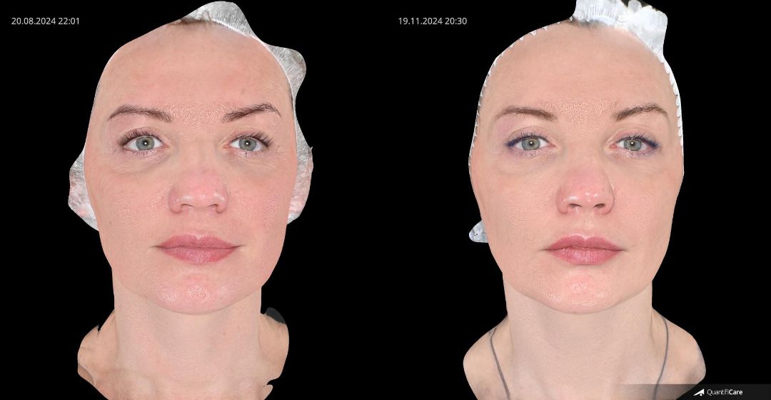
For modeling aesthetic outcomes. Real-time modeling visually demonstrates how expectations can be achieved, taking into account anatomical features. This mode shows whether more comprehensive anti-aging procedures are needed.
For assessing skin conditions:
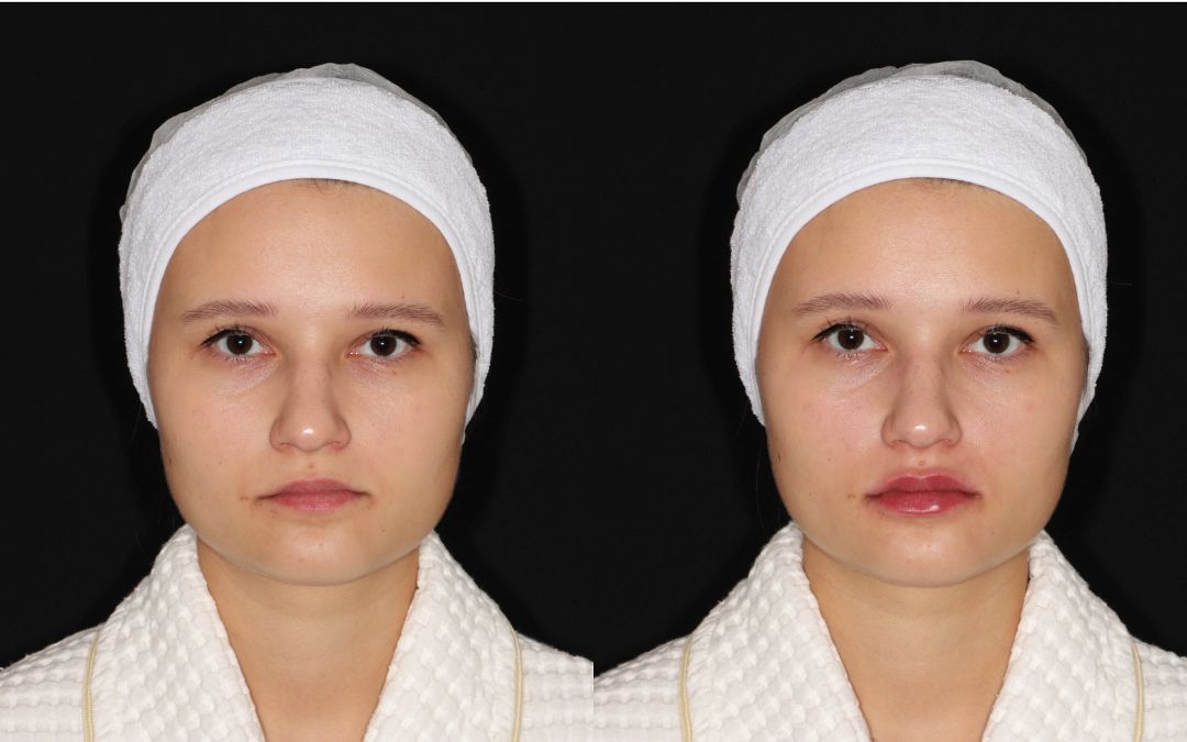
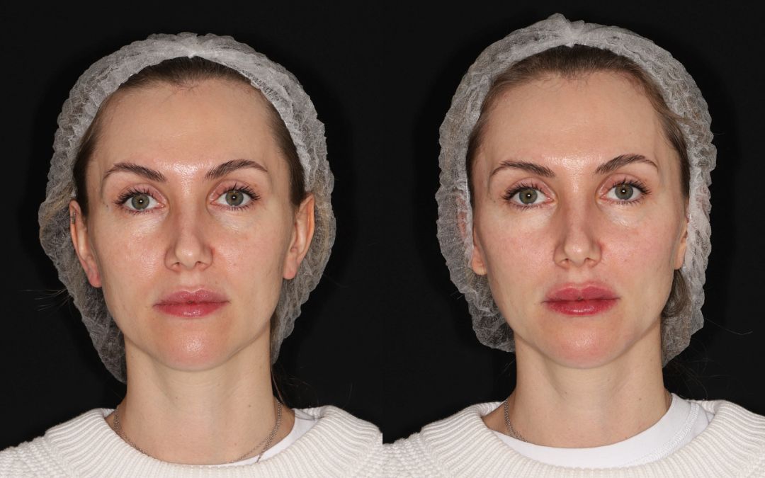
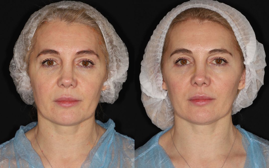
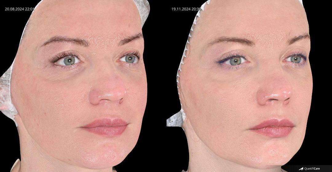
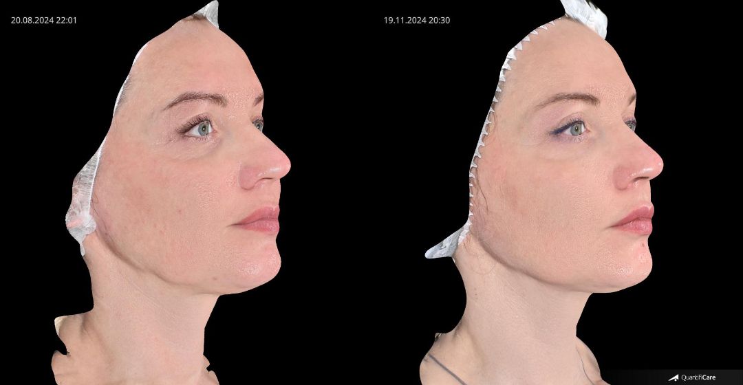
For clear "before/after" comparisons and "trying on" global beauty standards. We can visualize and digitize volume changes before and after procedures. We can measure angles, distances, and height-to-width proportions to check how close they are to the golden ratio.
Enables precise 3D measurements of anatomically localized areas in macro photography mode, such as scars, cellulite dimples, skin texture, wrinkle direction, and similar tasks. Alongside with general 3D face and body analysis, local 3D analysis of focal areas confirms the objectivity of results.
Quick 360° measurements and modeling. We can use the body contouring option for precise modeling and calculating expected volume changes.
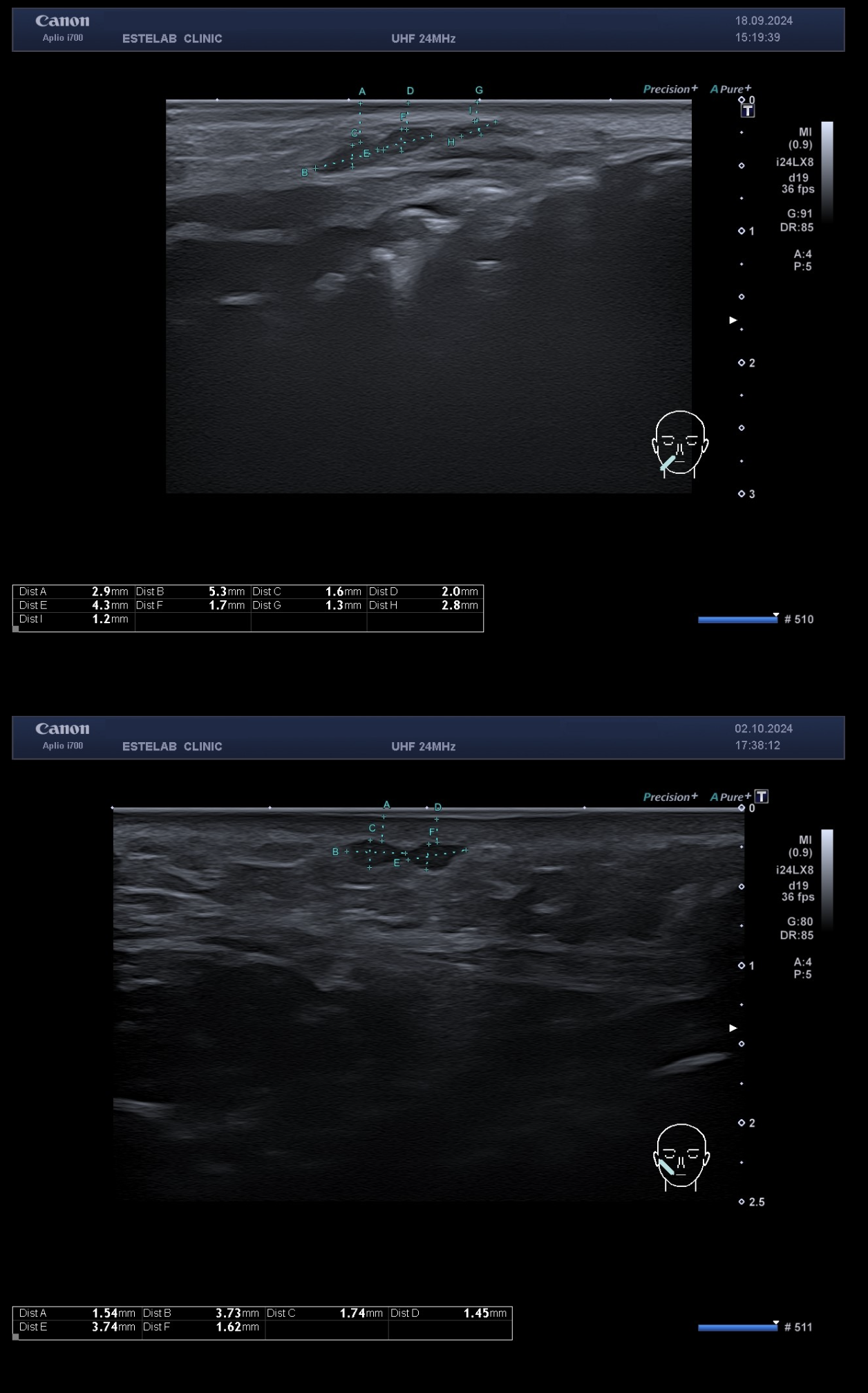
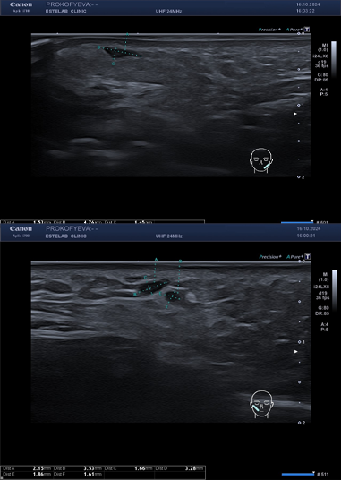
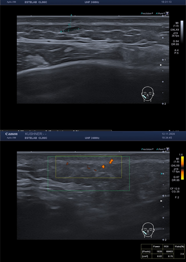
The ultrasound system supports ultra-wideband transducers, covering anatomical areas twice as large as conventional transducers. The system provides the best diagnostic quality in the shortest possible time.
Canon Medical Systems USA, Inc. introduces the industry's first ultra-high-frequency linear iDMS transducer with a 33 MHz frequency, delivering extremely precise near-field detail and the highest frequency on the market.
The new transducer expands the existing range of transducers currently available in the Aplio i-series ultrasound platform and is ideal for superficial subcutaneous imaging, superficial nerves, pediatric carotid artery studies, and more.
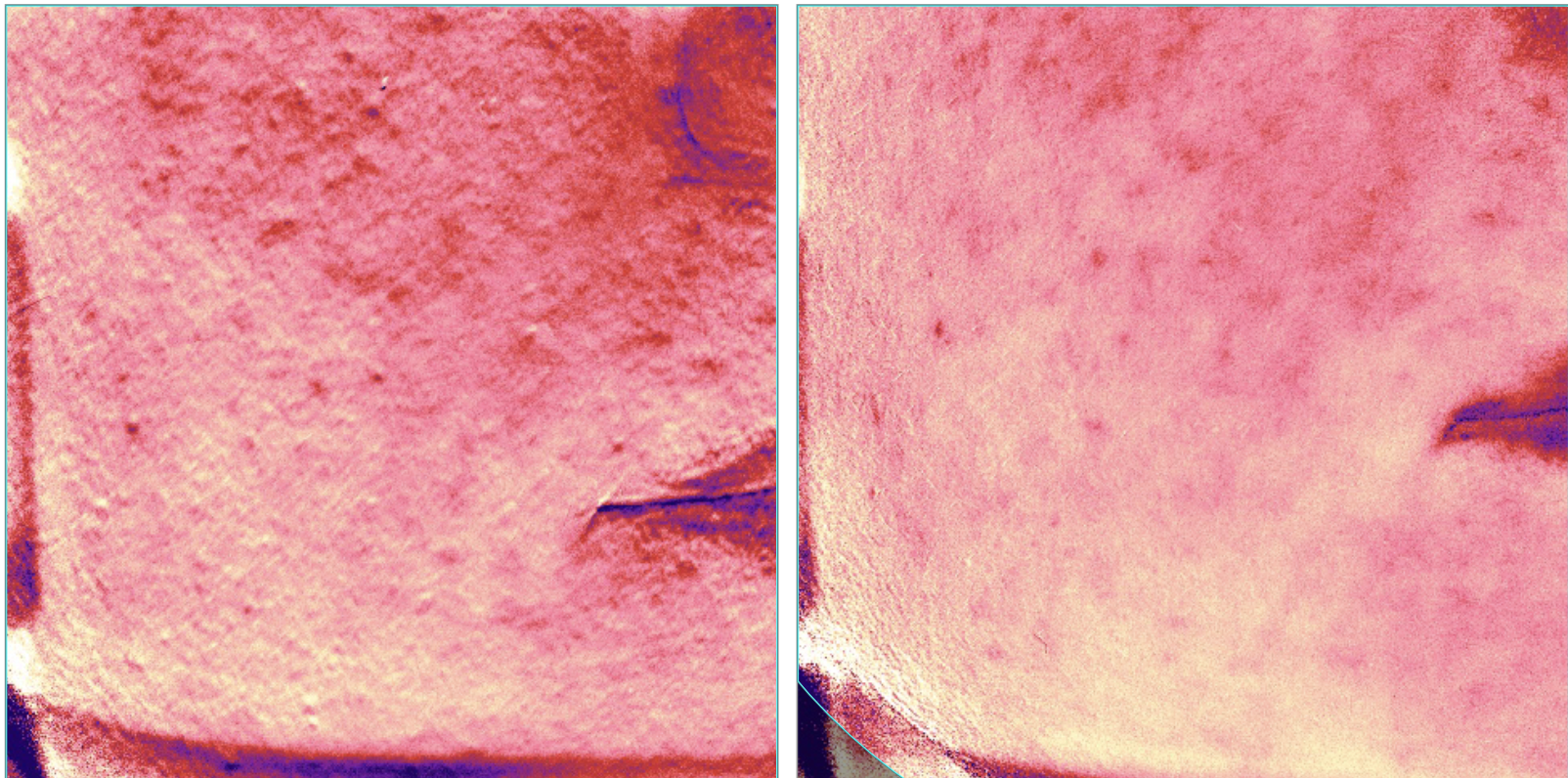
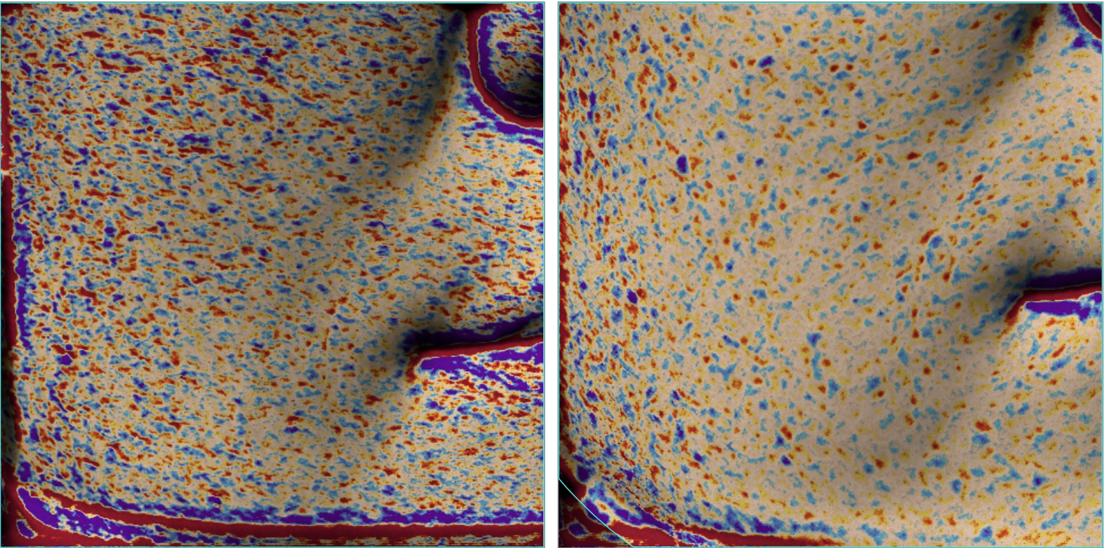
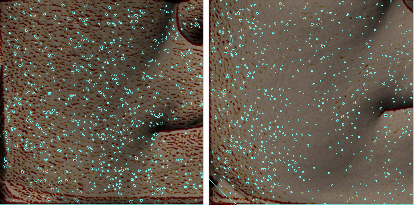
Antera 3D is a compact and easy-to-use device that allows for quick skin surface evaluation, creating and comparing computer-generated 3D skin images using multispectral analysis.
The Antera 3D system operates based on the interaction between light and biological tissues: incident light is partially absorbed and partially reflected by various skin structures, with these processes occurring at different intensities. The information captured during photography is converted into a 3D image on a computer monitor using specialized software. The system requires less than 90 seconds to perform a detailed analysis and generate a full report
The system uses diodes of different emission spectra, providing detailed information on:


Moscow, Bolshoy Levshinsky Lane, 6s1